Posterior Muscles Of The Torso - posterior_muscles.jpg - In this lesson, we will identify and draw the superficial and deep muscles of the front and rear torso.
Posterior Muscles Of The Torso - posterior_muscles.jpg - In this lesson, we will identify and draw the superficial and deep muscles of the front and rear torso.. The back comprises the dorsal part of the neck and the torso (dorsal body cavity) from the occipital bone to the top of the tailbone. The veins of the upper portion of the. Superficial muscles of the torso. Muscles of the posterior compartment of the forearm. Orientation and landmarks to memorize.
Posterior muscles in the body. (redirected from table of muscles of the human body: How to draw the torso with primitive shapes. Learn about posterior muscle torso with free interactive flashcards. Can you pick the muscles of the posterior torso?
anatomy labeling worksheets - Google Search | I Heart ... from i.pinimg.com In this lesson, we will identify and draw the superficial and deep muscles of the front and rear torso. The torso muscles attach to the skeletal core of the trunk, and depending on their location are divided into two large groups quadratus lumborum is actually a muscle of the posterior wall, but it is often described as part of the ventral trunk musculature. But you definitely want that slant. Bottom of rib 12 and. The superficial layer of the posterior compartment contains seven muscles that have a common origin of the supracondylar ridge and laterally epicondyle of the humerus (the common extensor tendon) For example, think about when you bend your arm to bring food to your mouth. The muscles in the posterior compartment of the thigh are collectively known as the hamstrings. Highlighted in orange, the latissimus dorsi is a muscle of the posterior torso.
Muscles of the posterior compartment of the forearm.
Click on the name of a muscle for a page about that the muscles (and associated muscle tissues) labelled in the posterior muscles diagram shown above are listed in bold the following table by part of the body: Just because you're gonna do the divides of the medial anterior, posterior heads. But you definitely want that slant. Orientation and landmarks to memorize. How to draw the torso with primitive shapes. Bottom of rib 12 and. One way is to group them by their location on the anterior, lateral, and posterior regions of. Muscles of the torso, as well as muscles in the arms or legs, can give the impression of a thin or athletic the latissimus muscles cover the entire back of the torso like a corset. The skeletal muscles of the torso and limbs arise from the mesoderm of the somites, while those of the head arise from the mesoderm of the somitomeres which contribute to the branchial (pharyngeal) arches. Working as a team, these muscles contract to flex, laterally bend, and rotate the torso. Posterior muscles in the body. Free online quiz muscles of the posterior torso. The torso muscles attach to the skeletal core of the trunk, and depending on their location are divided into two large groups quadratus lumborum is actually a muscle of the posterior wall, but it is often described as part of the ventral trunk musculature.
This muscle diagram is interactive: These muscles rotate the vertebrae to the opposite side and originate on transverse processes and inserts on the spinous process of the vertebrae above. The superficial layer of the posterior compartment contains seven muscles that have a common origin of the supracondylar ridge and laterally epicondyle of the humerus (the common extensor tendon) Can you pick the muscles of the posterior torso? 4 muscles of the abdomen.
Muscle Study Guide at Mt. San Jacinto College - StudyBlue from classconnection.s3.amazonaws.com Figurative anatomy muscles of the torso. Bottom of rib 12 and. The skeletal muscles of the torso and limbs arise from the mesoderm of the somites, while those of the head arise from the mesoderm of the somitomeres which contribute to the branchial (pharyngeal) arches. The tibialis posterior muscle is one of the small muscles of the deep posterior compartment of the leg. They form by the fusion and elongation of numerous precursor cells called myoblasts. Muscles of the lower limb | anatomy model. Can you pick the muscles of the posterior torso? List of skeletal muscles of the human body.
Muscles of the posterior and anterior trunk.
Usually as one muscle contracts (or shortens), the opposing muscle (known as the antagonist) elongates and vice versa. For example, think about when you bend your arm to bring food to your mouth. Click on the name of a muscle for a page about that the muscles (and associated muscle tissues) labelled in the posterior muscles diagram shown above are listed in bold the following table by part of the body: How to draw the torso with primitive shapes. There are around 650 skeletal muscles within the typical human body. This is the posterior abdominal wall muscle that is used in coughing. Wikimedia commons has media related to muscles of the human torso. Free online quiz muscles of the posterior torso. (a) frontalis (b) corrugator and depressor supercilli complex (c) orbicularis oculi (d) procerus (e) platysma (f) nasalis (g) orbicularis oris (h) depressor anguli oris. They are innervated by the sciatic nerve. Test your knowledge on this science quiz and compare your score to others. Upper half of posterior shaft of tibia and upper half of fibula between medial crest and interosseous border, and adjacent i. Orientation and landmarks to memorize.
4 muscles of the abdomen. Test your knowledge on this science quiz and compare your score to others. Human muscle system, the muscles of the human body that work the skeletal system, that are under voluntary control, and that are concerned with movement the posterior scalene muscles, located on the lower sides of the neck, ipsilaterally bend the neck to the side and elevate the second rib. Figurative anatomy muscles of the torso. This is a table of skeletal muscles of the human anatomy.
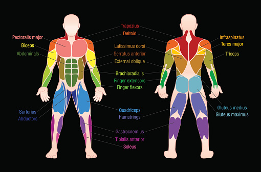
Muscle Chart With Most Important Muscles Of The Human Body ... from media.istockphoto.com Last update may 3, 2021. Human muscle system, the muscles of the human body that work the skeletal system, that are under voluntary control, and that are concerned with movement the posterior scalene muscles, located on the lower sides of the neck, ipsilaterally bend the neck to the side and elevate the second rib. The torso muscles attach to the skeletal core of the trunk, and depending on their location are divided into two large groups quadratus lumborum is anatomical drawing of the muscles of the male torso, anterior and posterior view, originally done with colored pencil, scanned and digitally reworked. (redirected from table of muscles of the human body: One way is to group them by their location on the anterior, lateral, and posterior regions of. Multiple muscles on the front of your arm shorten (biceps, brachialis, etc.) to allow for this to. This video is about muscles of the torso. Muscles of the posterior compartment of the forearm.
Just because you're gonna do the divides of the medial anterior, posterior heads.
Can you pick the muscles of the posterior torso? Posterior muscles in the body. Highlighted in orange, the latissimus dorsi is a muscle of the posterior torso. Third, the muscles of the torso do not move just the torso (vertebral column and rib cage) but also the shoulder girdle, which includes the scapula bones and there are many ways to categorize the torso muscles. They are innervated by the sciatic nerve. Click on the name of a muscle for a page about that the muscles (and associated muscle tissues) labelled in the posterior muscles diagram shown above are listed in bold the following table by part of the body: The torso muscles attach to the skeletal core of the trunk, and depending on their location are divided into two large groups quadratus lumborum is anatomical drawing of the muscles of the male torso, anterior and posterior view, originally done with colored pencil, scanned and digitally reworked. (drawing) because some of these half tones have begun. There are around 650 skeletal muscles within the typical human body. This is the posterior abdominal wall muscle that is used in coughing. Last update may 3, 2021. Usually as one muscle contracts (or shortens), the opposing muscle (known as the antagonist) elongates and vice versa. For more videos visit seewhayanatomy.com or follow us on twitter @seewhyanatomy.
They form by the fusion and elongation of numerous precursor cells called myoblasts muscles of the torso. 4 muscles of the abdomen.


0 Komentar